Retinal surgery
Prof. Dr. Abela-Formanek - Ophthalmologist for ophthalmology and optometry in Vienna
Retinal diseases
Retinal diseases can lead to serious vision loss and limitations in daily life.
The photo-sensitive retina is like a photographic paper where special cells in your retina react to light and pass signals to your brain that lets you see the world around you. The image is projected upside down on the retina and the image is erected in our head. The central retina or macula is responsible for color vision, facial recognition and reading. A functioning retina is important in order to be able to carry out complex movements such as hand-eye reflexes, grasping, catching, throwing and spatial orientation.
Damage to the central retina or visual center leads to loss of vision in the central visual field. Macular degeneration involves a slow, progressive loss of central vision caused by deposits in the retina. Other forms of macular degeneration arise from a cellophane-like membrane formation on the retina. This can be removed in certain cases. The surgery primarily serves to stabilize the disease and largely improve vision.
Focus on your eye health
Prof. Dr. Abela-Formanek looks back upon more than 25 years of experience in eye surgery and is considered an absolute expert in her field.
Epiretinal membrane (ERM) - Macula Pucker (MP)
Cause
The epiretinal membrane (ERM) is a thin cellular layer located on the surface of the retina. ERM arises from age-related changes in the eyes, but also from certain eye diseases such as diabetic retinopathy, veno-occlusive disease, after retinal surgery with retinal tears and laser treatment or even eye injuries. The risk of developing ERM increases with age.
Diagnosis
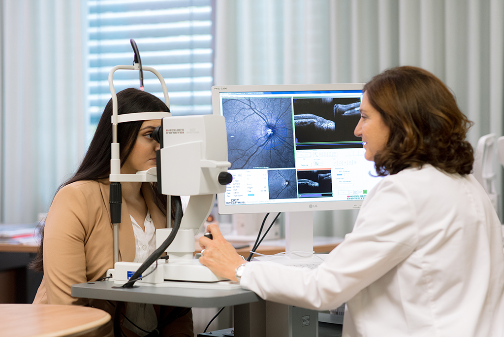
Optical coherence tomography (OCT) is used to image a slice of the retina to assess the severity of ERM.
Therapy and prognosis
In most cases, people with ERM do not need treatment as long as their vision is not severely impaired. However, if ERM progresses and affects vision, including the appearance of double vision and distortion, in most cases it will be necessary to perform a vitrectomy (removal of the vitreous and membranes). During this surgery, the ERM is removed from the retina using micro-forceps.
After a successful vitrectomy, there is an improvement in visual acuity and less distortion. However, it may take three to six months for vision to fully recover. The risks of this surgery are usually low, but a consultation is essential to establish the prognosis of the operation.
Retinal detachment
Retinal detachment is a serious eye problem in which the light-sensitive inner layer of the retina separates from its normal position. This medical emergency requires immediate medical attention as delayed treatment may result in permanent visual impairment or even loss of sight. The most common cause of a retinal detachment is when a retinal tear occurs due to a vitreous detachment.
Symptoms
- Sudden loss of vision, which may appear as a "curtain" obscuring part of the field of vision.
- The sudden appearance of flashes, flickering, or vitreous opacities in the field of vision.
- Decreased central visual acuity.
Treatment
In most cases, surgical intervention will be required to reattach the retina. A vitrectomy with gas filling will most often be necessary. Less commonly, a scleral buckle is attached to the sclera.
Macular degeneration (AMD)
A distinction is made between a dry and wet form of macular degeneration. Dry macular degeneration is a slowly progressive degeneration of the central retina. This leads to a deposition of metabolic waste products under the retina and a slow degeneration of the photoneurons. In some cases this form can change into a wet form.
What is wet AMD?
Wet AMD (called exudative AMD or neovascular AMD) is a form of AMD that involves the formation of abnormal, leaky blood vessels in the macula. These abnormal vessels can drain fluid and blood under the retina, causing swelling and scarring and affecting vision. Intravitreal injections can help slow the progression of wet AMD and stabilize or, in some cases, even improve vision.
What are intravitreal injections?
Intravitreal injections are minimally invasive procedures in which medications are injected directly into the vitreous humor (Vitreus) of the eye. The vitreous humor is the gel-like filler that fills the inside of the eye. These injections allow medication to be delivered exactly where it is needed, namely to the retina and the abnormal blood vessels in the macula.
Medication for wet AMD
The most commonly used drugs for intravitreal injections for wet AMD are anti-VEGF (vascular endothelial growth factor) drugs. These medications inhibit the growth and leakage of the abnormal blood vessels in the macula, thereby slowing the progression of AMD. Treatment consists of multiple injections and regular OCT examinations.
Prognosis:
Timely treatment of wet macular pathology leads to stabilization and in many cases, healing of AMD.
OCT – Ocular Coherence Tomography
Heidelberg Engineering
Accurate imaging of the retinal layers allows retinal disease to be ruled out before cataract surgery. An accurate diagnosis of wet macular disease can be detected early before symptoms appear. This enables timely treatment of the retina to prevent permanent retinal damage.
Would you like to make an appointment or do you have questions about operations?
Give us a call. Dr. Abela-Formanek will be happy to answer your questions!


